Pages

Showing posts with label bone. Show all posts

October 15, 2013

Notes on Anatomy and Physiology: The Spinal Column

We need to consider the form of the spine, its construction, if we are to eventually address two questions raised in the last note: what lengthens as we relax our weight, and how does the resulting separation of tissue promote health?
Accordingly, the next few posts will look at the various elements of the spine – the vertebral column as a whole, the individual vertebrae and the intervertebral discs, facet joints, spinal ligaments and thoracolumbar fascia. Take your time as you review these. Revisit them from time to time. Mulling over anatomy leads to unexpected insights into movement. And informs our understanding of the implications of illness.
Today, we will start with the spinal columnLying as it does near the middle of the body, it acts as an articulated, central pillar about which the rest of the body drapes and moves. The centrality of the spine and its vital connections with every corner of the body is beautifully expressed in Robert Frost’s poem, The Silken Tent:
She is as in a field a silken tent
At midday when a sunny summer breeze
Has dried the dew and all its ropes relent,
So that in guys it gently sways at ease,
And its supporting central cedar pole,
That is its pinnacle to heavenward
And signifies the sureness of the soul,
Seems to owe naught to any single cord,
But strictly held by none, is loosely bound
By countless silken ties of love and thought
To everything on earth the compass round,
And only by one’s going slightly taut
In the capriciousness of summer air
Is of the slightest bondage made aware.¹
Made up of 24 small, mobile bones called vertebrae and 9 fused sacral and coccygeal vertebrae, the spine appears straight when viewed from the front or back. Only when we look from the side

do we see that there are 4 reciprocal curves:
  • the cervical curve formed by the 7 cervical vertebrae. Concave posteriorly, it supports the head.
  • the thoracic curve made up of 12 thoracic vertebrae. This curve is convex posteriorly, thus making room for the heart, lungs and great vessels that lie within the chest.
  • the lumbar curve that lies fully in the centre of the abdominal cavity. It consists of 5 lumbar vertebrae and is concave posteriorly.
  • the sacral curve formed by 5 fused sacral and 4 fused coccygeal vertebrae. Convex posteriorly, it provides space for the organs found in the pelvis.
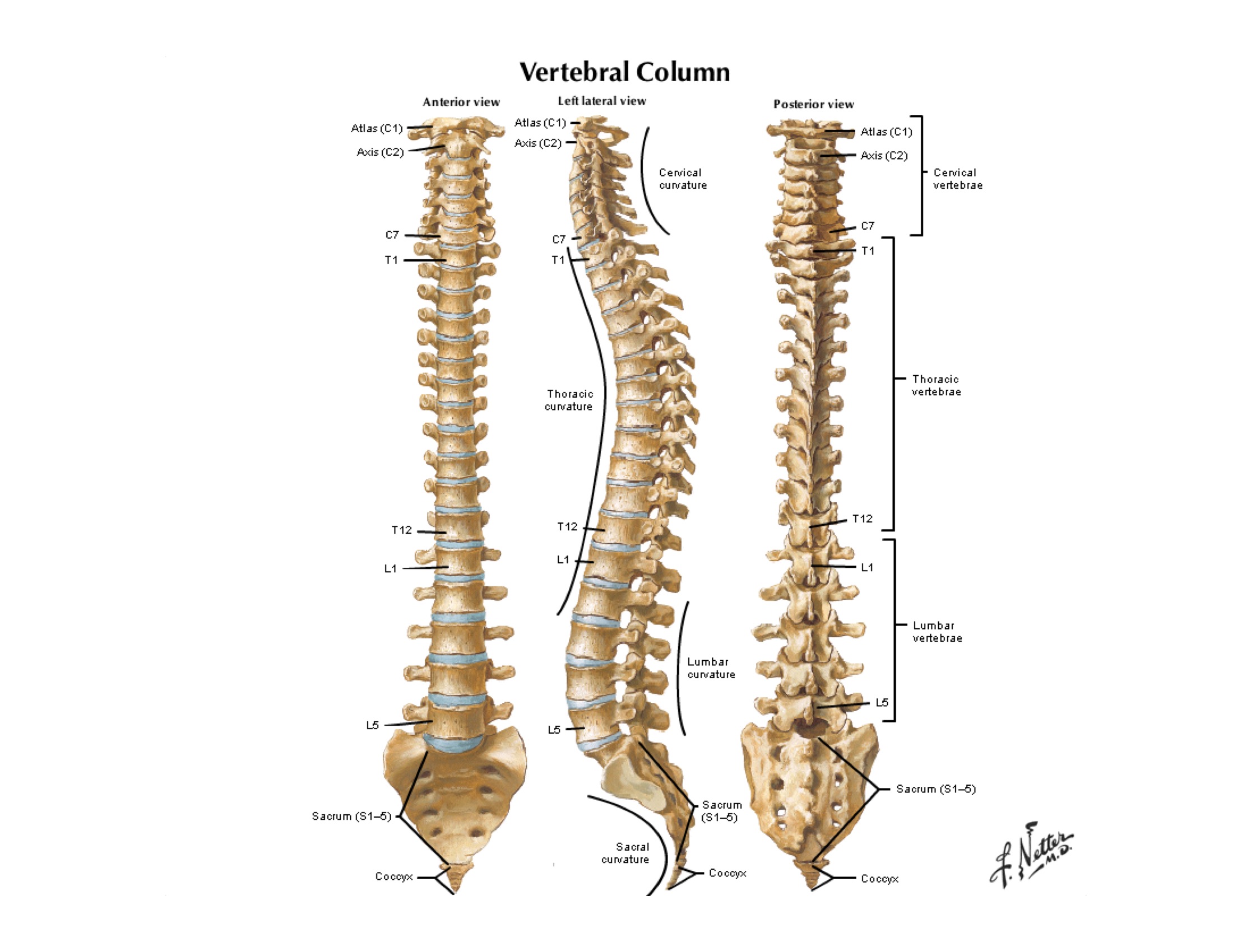
Fig 1 The curves of the spinal column are visible when seen from the side. Netter, 2006, Plate 153
Although the sacral curve is fixed, the other three are dynamic and change shape as we alter our posture and movements. Their flexibility helps the spine resist compression along its vertical axis by allowing the soft tissues that lie along the convex side of each curve to stretch and absorb some of the force.
As you watch others practice this coming week, notice how the don yu and tor yu play with these curves.
A number of factors produce these normal curves: the shape of the vertebral bodies and discs, the pull of ligaments and muscles, and the orientation of the facet joints. Disease, trauma and aging can push these curves out of their normal shape. Alter the curves enough and you risk straining the soft tissues, bones and joints of the spine. You also threaten the function of the nerve roots exiting the spine and crowd the space available for our organs.
You will remember from earlier posts that the spine, along with the skull and ribs, forms the axial skeleton – that part of our anatomy that surrounds and creates the spaces known as the dorsal and ventral cavities.

Fig 2 The axial skeleton, the centre pole of the body, viewed from the back and depicted in blue. Neumann, 2010, page 308

Fig 3 The axial skeleton, again shown in blue, as it looks from the front. Neumann, 2010, page 308.
We have already seen that the dorsal cavity houses the sensitive nervous tissue of the brain and spinal cord – tissue essential to life. And it is the ventral cavity that provides room for the other, equally vital, organ systems of the body.

Fig 4 Organs of the chest, abdomen and pelvis all lie within the ventral cavity created by the spine, ribs, pelvis and the soft tissues of the abdominal wall. Netter, 2006, Plate 548
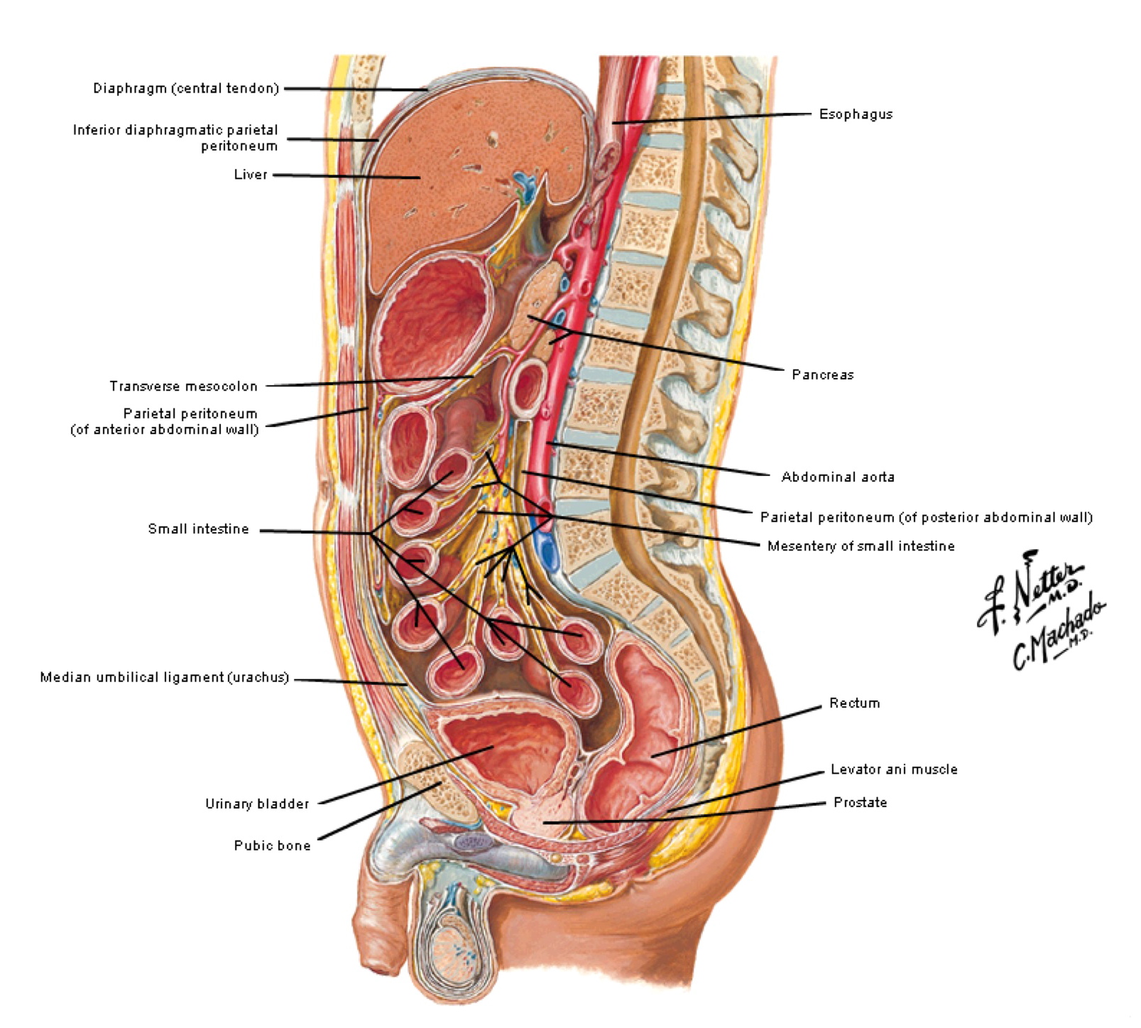
Fig 5 A picture with lots of detail. For now, just notice that this portion of the ventral cavity, bounded by the abdominal wall in front and the spinal column behind, is responsible for digestion, absorption of essential fluid and nutrients, reproduction and elimination. Netter, 2006, Plate 348
So, the spinal column, the centre pole about which the body is draped and moves. Constructed of many moving parts and capable of changing shape. Connected to all regions of the body.
With the next post, we turn to one of the building blocks of this column: the vertebrae and their discs.
¹ A Silken Tent taken from The Poetry of Robert Frost, Edward Connery Lathem, editor, 1942
² Kinesiology of the Musculoskeletal System, Foundations for Rehabilitation, Donald A. Neumann, Second Edition, 2010, Mosby Elsevier, ISBN 978-0-323-03989-5
³ Atlas of Human Anatomy, Frank H. Netter, 4th Edition, 2006, Saunders Elsevier, ISBN-13:978-1-4160-3385-1
© 2010 Taoist Tai Chi Society of Canada

The Vertebral Column Or Spinal Column

Vertebral column is also called back bone or spine and encloses the spinal cord. It is a flexible,curved , vertical rod, and consists of a row of 33 movably articulated ring like bones called vertebrae. Between each of the two bones the space is supplemented by pads of fibro-cartilage called the intervertebral discs. The vertebrae are held together by ligaments which prevent their dislocation, but permit a degree of movementt, making the backbone flexible. The adult vertebral column measures 60-70 cm in length.
The vertebrae are grouped and named according to the region they occupy.
Seven cervical vertebrae form the neck or cervical region.
Twelve thoracic vertebrae form the back of the thorax or chest.
Five lumbar vertebrae form the lumbar region or loins.
Five sacral vertebrae form the sacrum.
Four coccygeal vetebrae form the coccyx or tail.
The vertebrae in the three upper regions remain separate or distinct throughout life, and are called the movable vertebrae. Those in the two lower regions, the sacrum and coccyx, are united in the adult to form two bones and are called, the fixed vertebrae.
structure of human spinal column
                                           Human Vertebral Column
With the exception of the first two cervical vertebrae, all the movable vertebrae have similar structure; A typical vertebra is a bony ring. Its hole is called the vertebral foramen. The front border of the vertebral foramen is very thick. It is known as the body or centrum. It is amphiplatyan, that is flat on the upper as well as the lower side.
The remaining boundary of the vertebral foramen is thin. It is termed as the vertebral arch. Each half of a vetebral arch has a vetically narrow side, the pedicel, and a broader hind part, the lamina. The two laminae meet in the midline of the back. The upper and lower margins of the pedicel have concavities called the vertebral notches. When vertebrae are articulated together, adjacent notches form apertures-- the intervertebral foramina, for the exit of the spinal nerves. The vertebral arch gives off processes to which the muscles are attached. The processes include amedian spinous process and paired articular processes and transverse processes. The spinous process projects back and often also downward from the junction of the laminae. The articular processes of the adjacent vertebrae meet to form synovial joints. They provide limited movement between vertebrae. The vertebral foramina of all the vertebrae when intact form a vertebral canal that encloses the spinal cord.
In between the adjacent vertebrae, there are elastic pads of fibrocartilage- the intervertebral discs. This provide mobility to the vertebrae, check undue friction and take up shocks. Displacement of intervertebral disc is called slip-disc and is dangerous.
The cervical vertebrae
The cervical vertebrae are the smallest of the bones, and except the first and the second, which are peculiar in shape, the cervical vertebrae possess the following characters in common. The first cervical vertebra is called atlas. It is almost ring like. It provides up and down or nodding movement to the skull on it. The second cervical vertebra is termed as axis. Its centrum bears an odontoid process, which allows side to side or turning movement to the atlas and skull together on it.
The bodies of other cervical vertebrae are small and oblong in shape broader from side to side than from backward. The neural arch is large. The spinous processses are divided or bifid terminally. The transverse processes are perforated by foramina for the passage of the vertebral arteries. Thus this important blood vessel is protected as it passes through the vulnerable region of the neck.
cervical vertebrae

axis and atlas in cervical vertebrae

The Thoracic Vertebrae

Back to Top
These are 12 in number and are larger than the cervical vertebrae and they increase in size as they extend downwards. A typical thoracic vertebra has a heart-shaped body with facets on each for attachmentt of the ribs. The neural arch is relatively small, the spinous process is long and is directed downwards, and the transverse processses which help to support the ribs are thick and strong and carry articular facets for the ribs.
examples for thoracic vertebrae
Thoracic Vertebrae

The lumbar vertebrae

Back to Top
These are 5 in number and are located in the abdomen. These are the largest vertebrae consisting of a very large body which are kidney shaped. The spinous process is broad and hatchet-shaped. The transverse processes are long and slender. The fifth lumbar vertebra articulates with the sacrum at the lumbo-sacral joint.
lumbar vertebrae examples
Lumbar Vertebrae

The sacral vertebrae

Back to Top
These are 5 in number and are placed in the lower part of the vertebral column, wedged in between the two innominate bones and forming the back of the pelvic cavity.
human sacrum and coccyx
Human Sacrum and Coccyx

The coccygeal vertebrae

Back to Top
These are 4 in number and occur in the vestigial tail. They are very small, rudimentary and fused to form a curved, triangular bone, the coccyx or tail bone.

The curvature of the vertebral column

Back to Top
A notable feature in the vertebral column of human beings is the formation of four curves i.e., cervical, thoracic, lumbar and sacral, located in the neck, thorax, abdomen and pelvis respectively. The cervical and lumbar curves are directed forward and the thoracic and sacral curves are directed backward. Because of these curves, the centre of gravity is near the heels. This helps to maintain balance and makes walking erect, on two legs, much easier.
curvature of the vertebral column
Curvature of the Vertebral Column

Functions of the vertebral column

Back to Top
Vertebral column serves the following functions:
  • It acts as a firm support to the body.
  • It strengthens the neck and the trunk for upright posture while standing and walking.
  • It supports the ribs laterally, receives the weight of the arms through the ribs and rests on the hip bone which transfers the weight of the body to the legs.
  • Neural canal encloses and protects the spinal cord.
  • It forms a rigid vertical column from which viscera are suspended by mesenteries in the body cavity.
  • It provides flexibility to the trunk by having articular surfaces and intervertebral discs.
  • The upper cervical vertebrae provide free movement to the head in all planes.

Vertebral column

Vertebral column

From Wikipedia, the free encyclopedia

Vertebral column of a goat
The vertebral column, also known as backbone or spine, is a bony structure found in Vertebrates. It is formed from the vertebrae.

Humans[edit]

In human anatomy, the vertebral column usually consists of 24 articulating vertebrae,[1] and nine fused vertebrae in the sacrumand the coccyx. It is situated in the dorsal aspect of the torso, separated by intervertebral discs. It houses and protects thespinal cord in its spinal canal, and hence is commonly called the spine, or simply backbone.
There are normally 33 vertebrae in humans, including the five that are fused to form the sacrum (the others are separated byintervertebral discs) and the four coccygeal bones that form the tailbone.
The upper three regions comprise the remaining 24, and are grouped under the names cervical (7 vertebrae), thoracic (12 vertebrae) and lumbar (5 vertebrae), according to the regions they occupy. This number is sometimes increased by an additional vertebra in one region, or it may be diminished in one region, the deficiency often being supplied by an additional vertebra in another. The number of cervical vertebrae is, however, very rarely increased or diminished.[citation needed]

Other animals[edit]

In animals, vertebrae are defined by the regions of the vertebral column they occur in. Cervical vertebrae are those in the neckarea. With exception of two sloth genera (Choloepus and Bradypus) and the manatee (Trichechus),[2] all mammals have seven cervical vertebrae.[3] In other vertebrates it can range from a single vertebra in amphibians, to as many as 25 in swans or 76 in the extinct plesiosaur Elasmosaurus. The dorsal vertebrae range from the bottom of the neck to the top of the pelvis. Dorsal vertebrae attached to ribs are called thoracic vertebrae, while those without ribs are called lumbar vertebrae. The sacral vertebrae are those in the pelvic region, and range from one in amphibians, to two in most birds and modern reptiles, or up to 3 to 5 in mammals. When multiple sacral vertebrae are fused into a single structure, it is called the sacrum. The synsacrum is a similar fused structure found in birds that is composed of the sacral, lumbar, and some of the thoracic and caudal vertebra, as well as the pelvic girdle. Caudal vertebrae compose the tail, and the final few can be fused into the pygostyle in birds, or into the coccygeal or tail bone inchimpanzees (and humans).

Structure of individual vertebrae[edit]

Individual vertebrae are composed of a centrum (body), arches protruding from the top and bottom of the centrum, and various processes projecting from the centrum and/or arches. An arch extending from the top of the centrum is called a neural arch, while the hemal arch or chevron is found underneath the centrum in the caudal (tail) vertebrae offish, most reptiles, some birds, some dinosaurs and some mammals with long tails. The vertebral processes can either give the structure rigidity, help them articulate with ribs, or serve as muscle attachment points. Common types are transverse process, diapophyses, parapophyses, and zygapophyses (both the cranial zygapophyses and the caudal zygapophyses).

Classification[edit]

The centra of the vertebra can be classified based upon the fusion of its elements. In aspidospondyly, bones such as the neural spine, the pleurocentrum and the intercentrum are separate ossifications. Fused elements, however, classify a vertebra as having holospondyly.
A vertebra can also be described in terms of the shape of the ends of the centra. Centra with flat ends are acoelous, like those in mammals. These flat ends of the centra are especially good at supporting and distributing compressive forces. Amphicoelous vertebra have centra with both ends concave. This shape is common in fish, where most motion is limited. Amphicoelous centra often are integrated with a full notochordProcoelous vertebrae are anteriorly concave and posteriorly convex. They are found in frogs and modern reptiles. Opisthocoelous vertebrae are the opposite, possessing anterior convexity and posterior concavity. They are found in salamanders, and in some non-avian dinosaurs.Heterocoelous vertebrae have saddle-shaped articular surfaces. This type of configuration is seen in turtles that retract their necks, and birds, because it permits extensive lateral and vertical flexion motion without stretching the nerve cord too extensively or wringing it about its long axis.

Fish and amphibians[edit]

The vertebrae of lobe-finned fishes consist of three discrete bony elements. The vertebral arch surrounds the spinal cord, and is of broadly similar form to that found in most other vertebrates. Just beneath the arch lies a small plate-like pleurocentrum, which protects the upper surface of the notochord, and below that, a larger arch-shaped intercentrum to protect the lower border. Both of these structures are embedded within a single cylindrical mass of cartilage. A similar arrangement was found in the primitive Labyrinthodonts, but in the evolutionary line that led to reptiles (and hence, also to mammals and birds), the intercentrum became partially or wholly replaced by an enlarged pleurocentrum, which in turn became the bony vertebral body.[4]

a vertebrae bone (diameter 5 mm) of a small fish
In most ray-finned fishes, including all teleosts, these two structures are fused with, and embedded within, a solid piece of bone superficially resembling the vertebral body of mammals. In living amphibians, there is simply a cylindrical piece of bone below the vertebral arch, with no trace of the separate elements present in the early tetrapods.[4]
In cartilagenous fish, such as sharks, the vertebrae consist of two cartilagenous tubes. The upper tube is formed from the vertebral arches, but also includes additional cartilagenous structures filling in the gaps between the vertebrae, and so enclosing the spinal cord in an essentially continuous sheath. The lower tube surrounds the notochord, and has a complex structure, often including multiple layers ofcalcification.[4]
Lampreys have vertebral arches, but nothing resembling the vertebral bodies found in all higher vertebrates. Even the arches are discontinuous, consisting of separate pieces of arch-shaped cartilage around the spinal cord in most parts of the body, changing to long strips of cartilage above and below in the tail region. Hagfishes lack a true vertebral column, and are therefore not properly considered vertebrates, but a few tiny neural arches are present in the tail.[4]

Amniotes[edit]

The general structure of human vertebrae is fairly typical of that found in mammalsreptiles, and birds. The shape of the vertebral body does, however, vary somewhat between different groups. In mammals, such as humans, it typically has flat upper and lower surfaces, while in reptiles the anterior surface commonly has a concave socket into which the expanded convex face of the next vertebral body fits. Even these patterns are only generalisations, however, and there may be variation in form of the vertebrae along the length of the spine even within a single species. Some unusual variations include the saddle-shaped sockets between the cervical vertebrae of birds and the presence of a narrow hollow canal running down the centre of the vertebral bodies of geckos and tuataras, containing a remnant of the notochord.[4]
Reptiles often retain the primitive intercentra, which are present as small crescent-shaped bony elements lying between the bodies of adjacent vertebrae; similar structures are often found in the caudal vertebrae of mammals. In the tail, these are attached to chevron-shaped bones called haemal arches, which attach below the base of the spine, and help to support the musculature. These latter bones are probably homologous with the ventral ribs of fish. The number of vertebrae in the spines of reptiles is highly variable, and may be several hundred in some species of snake.[4]
In birds, there is a variable number of cervical vertebrae, which often form the only truly flexible part of the spine. The thoracic vertebrae are partially fused, providing a solid brace for the wings during flight. The sacral vertebrae are fused with the lumbar vertebrae, and some thoracic and caudal vertebrae, to form a single structure, the synsacrum, which is thus of greater relative length than the sacrum of mammals. In living birds, the remaining caudal vertebrae are fused into a further bone, the pygostyle, for attachment of the tail feathers.[4]
Aside from the tail, the number of vertebrae in mammals is generally fairly constant. There are almost always seven cervical vertebrae (sloths and manatees are among the few exceptions), followed by around twenty or so further vertebrae, divided between the thoracic and lumbar forms, depending on the number of ribs. There are generally three to five vertebrae with the sacrum, and anything up to fifty caudal vertebrae.[4]

Dinosaurs[edit]

The vertebral column in dinosaurs consists of the cervical (neck), dorsal (back), sacral (hips), and caudal (tail) vertebrae. Dinosaur vertebrae possess features known aspleurocoels, which are hollow depressions on the lateral portions of the vertebrae, which served to decrease the weight of these bones without sacrificing strength. Pleurocoels are filled with air sacs, according to some researchers. The sauropod dinosaurs are known for their unusually long tails, which were anchored in some cases by over 50 caudal vertebrae. In many hadrosaur and theropod dinosaurs, the caudal vertebrae were reinforced by tendons. The presence of three or more sacral vertebrae, in association with the hip bones, is one of the defining characteristics of dinosaurs. The occipital condyle is a structure on the posterior part of a dinosaur's skull which articulates with the first cervical vertebra.[5]

References[edit]

  1. Jump up^ "vertebral column" at Dorland's Medical Dictionary
  2. Jump up^ "Sticking Their Necks out for Evolution: Why Sloths and Manatees Have Unusually Long (or Short) Necks"May 6th 2011. Science Daily. Retrieved 25 July 2013.
  3. Jump up^ Frietson Galis (1999). "Why do almost all mammals have seven cervical vertebrae? Developmental constraints, Hox genes and Cancer".Journal of experimental zoology 285 (1): 19–26. doi:10.1002/(SICI)1097-010X(19990415)285:1<19::aid-jez3>3.0.CO;2-ZPMID 10327647.
  4. Jump up to:a b c d e f g h Romer, Alfred Sherwood; Parsons, Thomas S. (1977). The Vertebrate Body. Philadelphia, PA: Holt-Saunders International. pp. 161–170. ISBN 0-03-910284-X.
  5. Jump up^ Martin, A.J. (2006). Introduction to the Study of Dinosaurs. Second Edition. Oxford, Blackwell Publishing. pg. 299-300. ISBN 1–4051–3413–5.